Posted January 22, 2025 in Fertility Blog & Information
12 minute read
Key Takeaways
- Time-lapse monitoring in IVF offers continuous observation of embryo development, providing real-time imaging that enhances embryo quality assessment without invasive procedures.
- Second, this technology improves embryo selection by recording key developmental milestones. Therefore, it enables better ranking and selection of the best embryos for transfer.
- Ultimately, time-lapse imaging increases IVF success rates and results in more healthy live births. It lowers the risk of selecting low-quality embryos, improving patient satisfaction all around.
- Recent advancements in time-lapse technology, including high-resolution imaging and AI integration, have improved the accuracy of embryo evaluations and reduced subjective bias in assessments.
- Today, IVF practices trust time-lapse monitoring to help them make better decisions based on data. This transition paves the way for more personalized fertility treatments and can increase the chance of success for single embryo transfers.
- Ethical considerations are paramount with embryo selection using cutting edge technology. Most importantly, they call for transparency, informed consent, and unbiased interpretation of data when it comes to the use of IVF procedures.
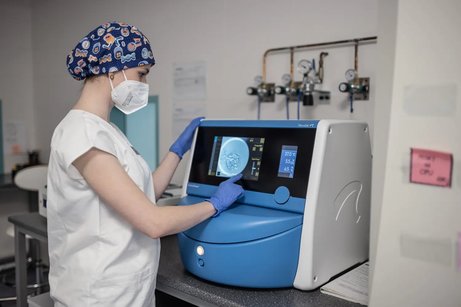
Time-lapse embryo monitoring in IVF provides a new perspective on how cutting-edge technology is improving the field of reproductive medicine. This approach provides a completely non-invasive and continuous imaging of embryos enabling embryologists to monitor development in great detail.
These observations give physicians the information they need to choose the most viable embryos to transfer. As a consequence, the overall in vitro fertilization success rates might improve dramatically. By offering a more complete view of embryo development, time-lapse monitoring helps you pinpoint critical developmental milestones to support more informed decision-making.
The widespread use of this technology would be a more equitable and effective step toward the shared aim of all – improving fertility outcomes. In turn, couples and individuals who choose to go through IVF can expect greater success rates with successful pregnancies.
Understanding this emerging technology can help illuminate how time-lapse embryo monitoring works and how it fits into the future of IVF.
What is Time-Lapse Monitoring
Time-lapse monitoring is a technique that has gained traction in recent years for closely monitoring embryo development. Opposed to conventional methods, including every now and then inspections, time-lapse systems allow steady imaging and evaluation of embryo quality.
This method allows researchers to capture important developmental milestones without disrupting the embryo’s environment. It provides a major benefit by enabling embryologists to choose only the embryos that are most likely to implant. This technology is non-invasive, allowing for embryos to remain within stable incubation environments. As a result, it can contribute to improved clinical outcomes.
Definition and Overview
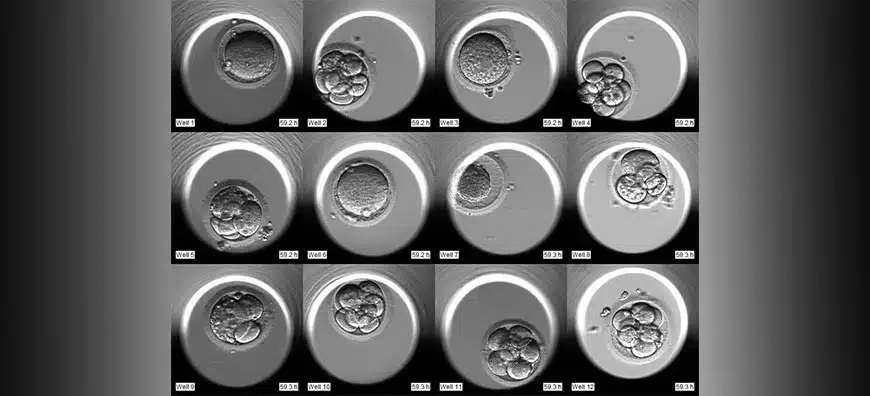
Time-lapse systems are unique from traditional approaches in that they enable uninterrupted observation from fertilization through to transfer. They’re a powerful tool because they document incredible, monumental developmental moments.
For instance, 58% of the embryos we monitored made it to the 4-cell stage by day 2, a significant milestone. This new technology has come a long way historically. It now boasts cutting-edge features such as the EEVA system, which predicts embryo viability at an early stage and improves embryologists’ selection process.
How It Works in IVF
This process requires rapid continuous imaging with a single low-intensity green LED illuminating multiple parallel focal planes. Such ongoing monitoring would allow the identification of embryos most likely to develop improperly and allow for interventions to be made in time.
Morphokinetic algorithms track embryo behavior during development, increasing selection accuracy and improving overall IVF efficiency. Such improvements have demonstrated ability to improve clinic pregnancy rates by more than 30%.
Benefits of Time-Lapse Imaging
Improved Embryo Selection
Time-lapse monitoring changes the game by allowing embryologists to monitor embryos around the clock, capturing footage of each embryo’s development. This technology is instrumental to both ranking embryos and understanding their development patterns. By tracking these distinct patterns, we are able to identify which embryos have the highest likelihood of a successful implantation.
Embryos that in the past would not have been considered are now recognized as good candidates for transfer. Time-lapse imaging uses a lot of morphokinetic parameters. These parameters allow for embryo scoring to be done in a much more efficient and accurate manner.
These parameters provide an unbiased snapshot of how the embryo is developing. For one, they’ve been shown to more accurately predict embryo quality—resulting in increased live birth rates—33.7% in the group that used time-lapse imaging.
Enhanced IVF Success Rates
Statistics highlight the positive impact time-lapse technology has had, even documenting an increase in live birth rates significantly among women aged over 38. By monitoring continuously, the chance of picking a bad embryo is minimized, resulting in only the best candidates being selected for transfer.
Time-lapse monitoring has been proven to be strongly correlated with ongoing pregnancy rates. In reality, the implantation rate in the time-lapse group was a whopping 81.3%. Similarly, the technology can predict aneuploidy in embryos without the need for invasive preimplantation genetic testing.
This feature alone would save millions in unnecessary procedures and increase patient happiness. The predictive power for identifying fetal heart pregnancy from these short videos is remarkable. With an AUC of 0.93, this method shows exceptional accuracy.
Advancements in Time-Lapse Technology
Recent developments in time-lapse imaging technology have fundamentally changed how embryos are tracked and managed in the in vitro fertilization (IVF) laboratory. These cultural advances made it possible to move the focus away from short-term profit and toward long-term culture. Therefore, they improve prediction of embryo quality and implantation potential.
High-resolution imaging has become an essential part of that process. Systems such as EmbryoScope, used by Care Fertility up until December 2022, take regular detailed images that are essential for determining which embryos to transfer. State-of-the-art lighting and camera technology beautifully captures these fleeting moments.
This enhanced imaging capability permits accurate detection of potentially life-threatening developmental defects and a much higher success rate.
Image Quality Improvements
With improved image quality, a clearer, more detailed picture of embryos can be captured leading to more accurate evaluations. Advanced lighting technology and high-speed cameras are central to this process. It guarantees that we document each important progress stage.
High-quality images are essential to spotting developmental defects early on, which in turn can improve the success rates of IVF procedures. Improved decision-making in selecting embryos for transfer depends on this definite identification.
We have clear evidence that applying morphokinetic scores increases pregnancy rates.
Integration with AI Tools
Artificial intelligence further optimizes embryo selection by analyzing large datasets efficiently. AI reduces subjective bias in assessments, offering objective insights into embryo viability.
The ability to process vast amounts of data enables AI to enhance predictive accuracy, despite the traditionally low predictive power of morphokinetic variables. This integration marks a significant step forward in IVF, promising more reliable outcomes for hopeful parents.
Impact on Embryo Selection
1. Criteria for Selection
Time-lapse embryo monitoring has revolutionized embryo selection, by eliminating the need to rely on subjective morphological characteristics and instead selecting embryos based on precise morphokinetic parameters.
Key factors include:
- Early cleavage stages are crucial, with early events like first cytokinesis (P1) and times between subsequent divisions (P2, P3) offering predictive insights into embryo viability.
- The speed at which an embryo reaches the blastocyst stage can indicate its implantation potential.
- High-quality blastocysts are more likely to result in successful pregnancies, as shown in predictive models by Van Royen et al.
- Synchronized development suggests a healthy embryo, increasing its chances of successful transfer and implantation.
2. Monitoring Embryo Development
The ability to track embryos in real-time is crucial for ensuring the best outcomes possible.
With continuous observation, embryologists can choose only the most viable embryos to transfer by recording each step of development.
Digital cameras take photos every 15 seconds, and these are later made into time-lapse videos.
These videos provide a clear view of embryonic development from 1 to 5–6 days of growth.
This approach increases the accuracy in selecting the embryos with the best implantation potential, ultimately translating to improved clinical outcomes.
3. Reducing Human Error
Automated monitoring minimizes subjective errors, offering consistent data collection that surpasses manual evaluations.
This objective approach enhances selection accuracy, with studies showing higher ongoing pregnancy rates in time-lapse monitored groups compared to traditional methods.
The integration of technologies like preimplantation genetic screening further refines embryo assessment, maximizing implantation success.
Time-Lapse Monitoring Process
Understanding the details of time-lapse embryo monitoring in IVF starts with properly configuring your equipment. This includes making sure the time-lapse incubators are functional with the lab equipment already in use, as well as calibrating the incubators to run smoothly and accurately.
Creating a stable environment for embryos to develop is essential. Use low-intensity red LED illumination at 635 nm and provide less than 0.5 s exposure per image. This low intensity illumination allows for long-term monitoring, imaging the embryo development every few minutes without affecting the culture.
Equipment and Setup
| Incubator Type | Features | Price Range | Pros | Cons | Compatibility |
|---|---|---|---|---|---|
| Basic Model | Standard imaging | $5,000-$10,000 | Affordable | Limited features | High |
| Advanced Model | Dark field illumination | $15,000-$20,000 | High accuracy | Costly | Medium |
| Premium Model | Kinetic analysis | $25,000+ | Best performance | Expensive | High |
Data Collection Methods
Advanced software is available to organize and interpret imagery collected via time-lapse monitoring. This software allows for real-time monitoring of important kinetic parameters.
For instance, it tracks cell cycle time, which is critical for determining which embryos are optimal to implant. The system increases the rate of pregnancy by 36%.
Its main strength lies in its predictive ability to determine which embryos are likely to fail upon implantation. By capturing very specific intervals of time, we’re able to analyze embryo development with a new level of detail, and extreme quality follows.
Implications for IVF Practices
Changes in Transfer Practices
Time-lapse embryo monitoring technology is providing IVF clinics with a new approach to debunking standard embryo transfer protocols. With uninterrupted information about embryo growth, it can provide accurate information about the best time to make transfers.
It provides for continuous observation of embryos which enables clinicians to identify the perfect time for embryo transfer. This strategy might provide an impressive increase in probability of successful implantation.
Thanks to time-lapse imaging, the selection of embryos for transfer is now more informed, allowing for higher accuracy in choosing those with better developmental prospects. This technique has dramatically reduced the risk of multiple gestation pregnancies.
It incentivizes single embryo transfers, creating assurance that the best embryo is selected.
Ethical Considerations
The introduction of advanced technology such as time-lapse monitoring to embryo selection raises ethical questions to the forefront. While it improves embryo selection, it can bring in new biases by how data is interpreted.
Most importantly, it’s imperative that IVF practices are transparent, providing patients with the information they need to make informed decisions and consent to using the technology. Research outlines significant advantages, such as increased clinical pregnancy rates and greater birth weights.
These findings highlight the urgent need for ethical scrutiny. Even with these improvements, technology should never replace the patient’s ability to make an informed decision.
Most importantly, it’s important to make sure we aren’t treating anyone unfairly.
Conclusion
It accelerates embryo selection, providing physicians with a better understanding of embryo development without interrupting growth. Using this technology, clinics can maximize success rates by selecting only the most viable embryos. Patients feel less anxious, assured in the knowledge that the process is more accurate and data-driven. For anyone currently traveling along an IVF journey, time-lapse embryo monitoring is a seamless integration of technology and compassion, providing both hope and understanding.
Take full advantage of these new improvements to get the most out of your IVF journey. If you’re a patient or a practitioner, being aware of these evidence-based methods can be transformative. Explore the additional benefits time-lapse monitoring can offer and how it can help you achieve your parenthood dreams.
Frequently Asked Questions
What is time-lapse monitoring in IVF?
Time-lapse monitoring is a technology that fertility clinics around the world are increasingly adopting. It enables embryologists to observe embryo behavior and developmental patterns without being invasive, thereby improving the accuracy of embryo selection and increasing the chances of successful pregnancy.
How does time-lapse imaging benefit IVF?
As time-lapse imaging provides continuous embryo observation, it minimizes the need to physically handle embryos. Second, it enhances embryo selection accuracy, thanks to comprehensive growth information, increasing the likelihood of successful implantation rates.
What are the advancements in time-lapse technology?
Recent developments have introduced enhanced image resolution and AI capabilities, significantly improving time lapse embryo monitoring systems. These advancements provide new capabilities for unprecedented analysis of embryo development, playing an important role in achieving the best success rates possible across all fertility treatments.
How does time-lapse monitoring impact embryo selection?
With a time-lapse embryo monitoring system, you gain detailed and continuous developmental data, significantly enhancing embryologists’ ability to select viable embryos, thus optimizing the chances for successful pregnancy outcomes through IVF cycles.
What is involved in the time-lapse monitoring process?
The process consists of putting embryos into a specific incubator with cameras. Each embryo is imaged at predetermined intervals. This provides the clinic with a time-lapse video sequence of individual embryo development, which can help inform selection decisions.
How has time-lapse monitoring changed IVF practices?
Time-lapse embryo monitoring has transformed IVF by providing continuous embryo observation and more accurate information. This advanced technique enhances embryo selection and minimizes the risk of handling human embryos, ultimately resulting in improved outcomes in IVF clinics globally.
Are there any implications for IVF practices with time-lapse monitoring?
Yes, time-lapse monitoring improves embryo assessment accuracy. It supports better decision-making in embryo selection. This can lead to higher success rates and more efficient IVF treatments.